Cell culture assays identify histatins as the major wound-closure stimulating factors in human saliva, distinguishing oral mucosal repair from cutaneous healing through specific peptide-driven mechanisms rather than systemic growth factor cascades. Oudhoff et al. established this functional dichotomy by isolating histatins as the primary active components in salivary wound closure assays, separating their activity from serum-derived growth factors in FASEB Journal.
These biochemical pathways explain the rapid re-epithelialization observed in oral tissues compared to skin, yet current evidence remains restricted to preclinical models and cell lines. No completed human trials have validated synthetic histatin formulations for therapeutic use in wound treatment or antifungal defense.
While peptides for healing in systemic contexts often rely on growth hormone secretagogues, salivary histatins operate through innate immune modulation and direct cell-matrix interactions that lack validated pharmacokinetic data linking physiological concentrations to therapeutic efficacy.
Integrin Signaling and Angiogenic Limits
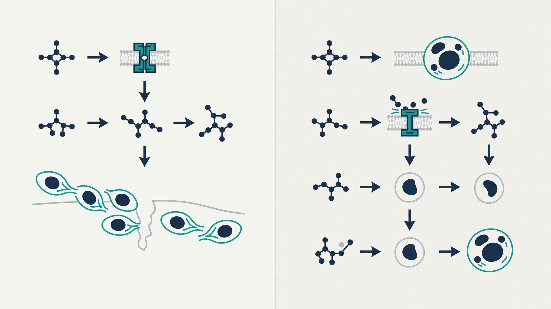
Figure 1: Histatin-1 integrin signaling and non-lytic antifungal mechanisms in oral wound repair.
Histatin-1 drives re-epithelialization through specific molecular interactions with cell surface receptors rather than generic mitogenic stimulation. In vitro wound closure and Boyden chamber assays demonstrate that Histatin-1 promotes endothelial cell adhesion and spreading specifically on fibronectin substrates. This activity depends on integrin binding and the activation of the MAPK/ERK signaling cascade.
Human primary cultured endothelial cells and the EA.hy926 cell line show enhanced migration when exposed to Histatin-1, but this effect is abolished when integrin function is blocked.
The peptide facilitates motility through cytoskeletal reorganization rather than simple proliferation, a distinction that matters for understanding scarless healing versus fibrotic repair. Torres et al. confirmed in Oral Diseases that this activity is intrinsic to salivary biochemistry rather than a secondary effect of blood contamination, identifying histatins as the dominant closure factors in cell culture.
Angiogenesis claims regarding Histatin-1 require strict qualification based on the available evidence hierarchy. Chick chorioallantoic membrane models and in vitro tube formation assays indicate that Histatin-1 promotes vascular structure formation in non-mammalian and isolated cell systems. These findings suggest a potential role in the proliferative phase of healing where neovascularization supports granulation tissue, but mammalian in vivo vascular data are lacking.
The chick embryo model provides preliminary evidence of bioactivity but does not replicate human oral mucosal physiology, immune responses, or hemodynamic forces. Researchers must distinguish between avian developmental angiogenesis and adult human wound neovascularization when interpreting these results. Regulatory standards for claiming angiogenic efficacy in humans require robust mammalian data that do not currently exist for this peptide, limiting current findings to mechanistic hypotheses rather than clinical endpoints.
The biochemical environment of the oral cavity amplifies these peptide effects in ways that are difficult to replicate synthetically. Salivary histatins stabilize fibrin matrices through interactions with proline-rich proteins and statherin, facilitating early cell adhesion and migration. This stabilization occurs faster than cutaneous coagulation processes, creating a provisional matrix optimized for rapid closure.
Oral mucosal macrophages transition from pro-inflammatory M1 to pro-resolving M2 phenotypes more rapidly than their cutaneous counterparts, a process supported by salivary bioactive compounds including histatins, mucins, and growth factors like EGF and VEGF. Despite robust in vitro characterization, the distance to clinical application persists. Unlike peptides for joint and tendon healing, which have entered clinical evaluation for musculoskeletal indications with defined endpoints, histatin research has not progressed to registrational trials for oral wounds.
Salivary extracellular vesicles have emerged as potential delivery vehicles in laboratory studies, but diagnostic applications currently outpace therapeutic development.
Mitochondrial Targeting and Delivery Engineering
Histatin-5 employs an antifungal mechanism fundamentally distinct from pore-forming antimicrobial peptides like LL-37 or defensins. Mitochondrial membrane permeabilization assays demonstrate that Histatin-5 translocates into Candida albicans cells and targets mitochondrial membranes without disrupting the plasma membrane. This intracellular trafficking leads to rapid, non-lytic release of intracellular ATP, causing cell death through energy depletion rather than the membrane lysis characteristic of many cationic antimicrobial peptides.
Pharmacological studies confirm that this ATP release is the primary lethal event.
Reactive oxygen species do not mediate this candidacidal activity, as biochemical exclusion assays show that ROS scavengers fail to protect C. albicans from Histatin-5 killing. This specificity matters for therapeutic design because agents relying on ROS generation often face host toxicity issues due to collateral damage to mammalian cells.
Histatin-5’s targeted mitochondrial disruption offers a theoretically safer profile, though selectivity for fungal over human mitochondria requires rigorous validation in mammalian cell systems before safety can be assumed.
Recent materials science research has attempted to leverage this mechanism for drug delivery in response to escalating antifungal resistance. Reyes-Torres et al. engineered experimental nanodevices functionalized with Histatin-5 as a stimuli-responsive gate, reporting enhanced antifungal activity against C. albicans in ACS Applied Materials & Interfaces. These mesoporous silica nanoparticles release surfactants upon Histatin-5 recognition of fungal targets, combining membrane disruption with the peptide’s native activity.
Serial passage assays indicate negligible resistance development with this dual-action strategy over multiple generations of exposure. However, this represents advanced material science rather than validated therapy. The nanodevice remains an experimental platform for overcoming antifungal resistance, not a clinical product. Current evidence characterizes this mechanism exclusively in C. albicans, and efficacy against other oral fungal pathogens remains unestablished in supplied records.
The leap from nanoparticle efficacy in a controlled assay to clinical management of oral candidiasis involves unresolved questions regarding mucosal penetration, retention time, and host safety.
Comparisons with other mucosal defense peptides highlight Histatin-5’s unique niche and limitations. Human beta defensins immunology research shows that defensins accelerate tissue repair through keratinocyte proliferation and angiogenin secretion alongside antimicrobial activity. LL-37 promotes wound healing but carries risks of pathological angiogenesis and inflammation with prolonged exposure, as demonstrated in rosacea models. Histatin-5 lacks these immunomodulatory side effects in current literature, focusing its bioactivity on fungal clearance and, to a lesser extent, endothelial migration.
This functional separation may reduce off-target risks but also limits the peptide’s utility as a broad-spectrum wound healer compared to multifunctional cathelicidins. The choice between these peptide classes for therapeutic development depends on the specific clinical indication: Histatin-5 for fungal control with minimal inflammation, defensins or LL-37 for broader tissue regeneration with higher risk profiles.
Clinical Validation and Regulatory Standards
The absence of clinical trial data represents the most significant barrier to histatin therapeutic adoption. A search of ClinicalTrials.gov identifies protocol NCT01078467 related to the molecular anatomy of oral wound healing, but no results demonstrating efficacy of topical histatin formulations for oral wounds appear in supplied records. This gap contrasts sharply with the extensive in vitro literature. Regulatory agencies require human safety and efficacy data for peptide therapeutics, regardless of mechanistic plausibility. The U.S.
FDA classifies wound dressings combined with drugs under specific regulatory frameworks that demand substantial equivalence or new drug applications, as outlined in FDA Executive Summary on Wound Dressings. Synthetic histatins would likely face scrutiny as new molecular entities requiring full drug development pathways rather than approval as natural supplements or medical devices.
Transatlantic regulatory divergence may influence development trajectories for these peptides. European frameworks for advanced therapy medicinal products offer potential pathways for complex biologicals, while U.S. regulations emphasize well-controlled trials with standardized endpoints. Neither jurisdiction has approved histatin-based wound products.
The European Medicines Agency evaluates antifungal efficacy through rigorous microbiological success criteria, as detailed in the Ecalta Scientific Discussion, setting a high bar for claims regarding Candida clearance that histatin therapeutics have not yet met in human studies.
Pharmacokinetic uncertainties compound regulatory challenges. Salivary biochemistry literature documents endogenous histatin concentrations, but no studies establish therapeutic thresholds in wound beds. The oral cavity presents a hostile environment for peptide stability. Proteolytic degradation, salivary flow, and swallowing mechanics may prevent synthetic histatins from achieving sustained bioactive concentrations at injury sites. Hydrogel and nanoparticle delivery systems show promise in vitro but lack human pharmacokinetic validation.
Long-term tissue outcomes also require investigation beyond initial closure metrics. Mouse hard palate injury models reveal that rapid re-epithelialization is followed by months of cellular remodeling that extends far beyond apparent wound closure. Histological and epigenetic analyses published in Cell Death & Disease show that epithelial homeostasis is re-established through prolonged molecular programs lasting up to six months post-injury. Whether histatin-driven acceleration of initial closure affects this extended remodeling phase remains unknown.
Rapid healing that compromises long-term barrier function or tissue architecture would be clinically counterproductive. Direct measurement of barrier permeability in healed tissue is needed to assess full functional restoration before regulatory approval can be considered. Researchers evaluating these peptides must maintain strict separation between in vitro mechanism and therapeutic claim, acknowledging that biochemical plausibility does not substitute for clinical evidence in the absence of registrational trials.
Further Clinical & Regulatory Context
For deeper analysis and cross-referenced evidence, see: - Related Clinical & Pharmacological Analysis: Tuftsin Macrophage Activation: Neuropilin-1 Kinetics vs Clinical Efficacy - Related Clinical & Pharmacological Analysis: Thymic Peptides in Immunosenescence: Distinguishing Regeneration from Expansion - Related Clinical & Pharmacological Analysis: Cyclic Antimicrobial Peptide Pharmacology: LPS Binding Versus Renal Uptake