
Tuftsin binds specifically to neuropilin-1 on macrophages with an equilibrium dissociation constant of 5.3 × 10⁻⁸ M, a kinetic parameter established in ligand-binding studies that correlates with subsequent phagocytosis stimulation in vitro (PubMed). This precise molecular interaction defines the biological plausibility of tuftsin peptide macrophage activation, yet it exists largely within controlled laboratory environments rather than systemic therapeutic applications.
The tetrapeptide’s capacity to enhance innate immune function is mechanistically sound at the receptor level but practically constrained by rapid proteolytic degradation that limits its utility as a standalone drug. Victor Najjar identified this endogenous activator in 1970 at Tufts University, isolating it from residues 289–292 of the IgG heavy chain CH2 domain according to historical records compiled by Peptide Insight.
Despite decades of investigation into this spleen-derived peptide, no completed Phase III trials have demonstrated clinical efficacy for tuftsin monotherapy in infection or malignancy within FDA or EMA registries. The disconnect between robust binding kinetics and absent clinical endpoints highlights a persistent challenge in translating short-lived endogenous peptides into stable pharmaceutical agents.
Neuropilin-1 Binding and Receptor Internalization
The functional claims surrounding tuftsin rest entirely on its stoichiometry with neuropilin-1 (Nrp1). Research confirms that the peptide does not merely associate with cell membranes but engages this specific receptor to trigger downstream signaling cascades. The reported Kd of 5.3 × 10⁻⁸ M indicates high-affinity binding essential for initiating receptor-mediated endocytosis, a process documented in foundational studies on macrophage internalization (PubMed).
Without this specific engagement, the tetrapeptide remains biologically inert regardless of concentration.
Binding alone does not equate to sustained activation. The peptide must induce conformational changes that facilitate cytoskeletal rearrangement for phagocytosis. Recent work on tuftsin-based fusion proteins demonstrates that retaining this Nrp1 binding capability is necessary for enhancing antitumor efficacy, specifically through CD47 down-regulation pathways. When the peptide is incorporated into larger constructs like Ec-LDP-TF, it significantly enhances macrophage phagocytic activity compared to controls lacking the tuftsin moiety.
This suggests that while the native tetrapeptide provides the critical targeting signal, its therapeutic value may only be realized when protected within a larger molecular scaffold.
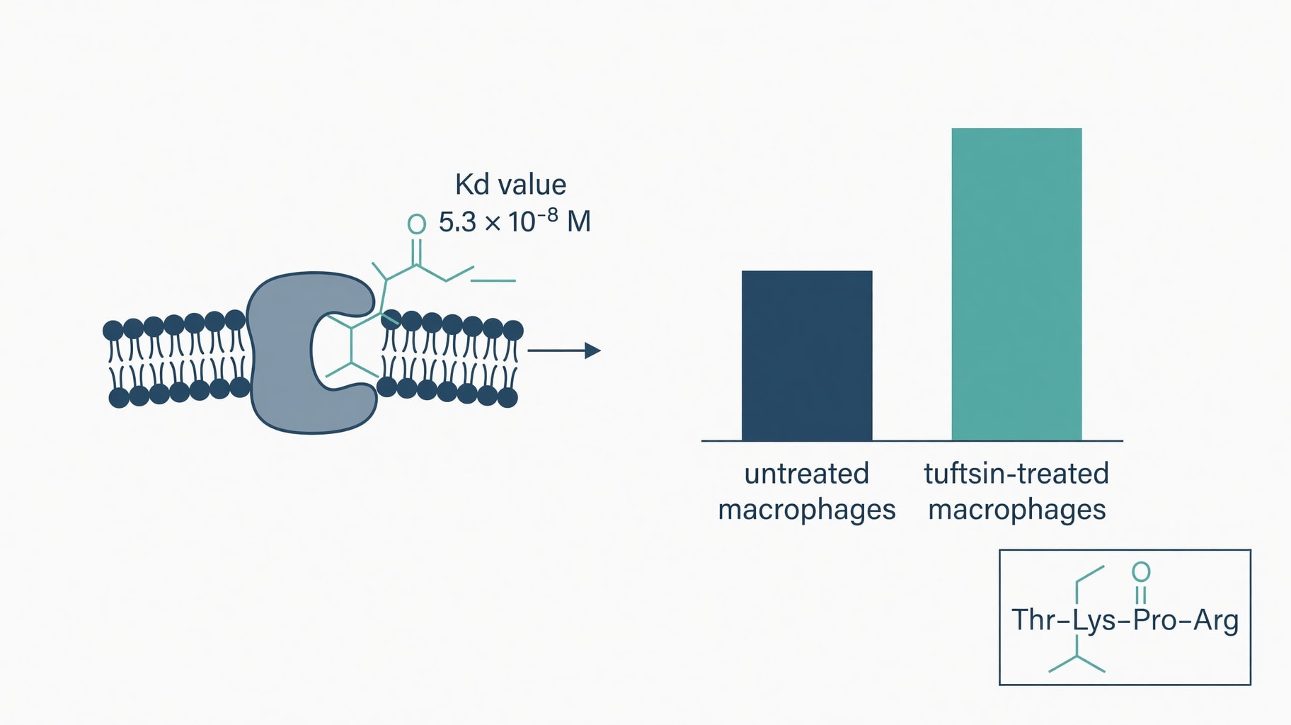
Figure 1: Tuftsin-neuropilin-1 binding kinetics and correlation to in vitro macrophage phagocytosis enhancement.
Signaling through neuropilin-1 also intersects with transforming growth factor beta pathways, as noted in mechanistic reviews of tuftsin signaling (PubMed). This crosstalk complicates the interpretation of isolated phagocytosis assays. Activation of Nrp1 can modulate microglial and macrophage phenotypes in ways that extend beyond simple particle ingestion.
Researchers must therefore distinguish between acute phagocytic stimulation and broader immunomodulatory effects that may alter disease outcomes in autoimmune or neoplastic contexts.
The specificity of the Kd measurement provides a necessary anchor, but the downstream functional consequences remain highly context-dependent. Furthermore, the distinction between tuftsin and other immune-modulating peptides is critical. While tuft cells in epithelial tissues also regulate phagocytosis and inflammation resolution through specialized pro-resolving mediators, they operate through distinct gustatory signaling proteins and transcription factors like Pou2f3, unrelated to the neuropilin-1 pathway utilized by the tuftsin tetrapeptide.
Conflating these distinct biological entities obscures the specific receptor-ligand kinetics that define tuftsin pharmacology.
Phagocytic Index Assays and Methodological Constraints
Quantifying tuftsin’s effect requires rigorous phagocytic index assays, yet reproducibility has historically plagued the field. Early difficulties in demonstrating consistent phagocytosis stimulation led to the development of specialized human polymorphonuclear leukocyte assays designed specifically for this peptide (PubMed). Standardized protocols are now essential because generic phagocytosis assays often fail to capture the subtle, concentration-dependent effects of native tuftsin.
Variability in donor cells, incubation times, and target particles can obscure genuine biological signals or generate false positives. Most positive data derives from murine peritoneal macrophages or human neutrophils in vitro. These models provide clean mechanistic readouts but poor predictive value for systemic human efficacy. In a living organism, the peptide faces immediate enzymatic destruction. The half-life of native tuftsin is measured in seconds according to comparative stability analyses.
This fleeting existence means that even if receptor affinity is optimal, the peptide may be degraded before achieving sufficient occupancy to drive a therapeutic response in vivo.
Modern assay development continues to refine these measurements using fluorescently labeled bioparticles and flow cytometry. Commercial platforms now combine surface marker profiling with functional readouts to characterize macrophage responses more comprehensively Cell-based Macrophage Assays. However, these advanced methods still primarily validate tool compounds or engineered fusion proteins rather than native tuftsin itself.
The methodological evolution reflects a shift from studying the natural peptide as a drug candidate to using it as a targeting ligand for more complex therapeutics.
For instance, nanoparticle systems responsive to tumor-associated proteases like legumain can release active tuftsin fragments specifically within the tumor microenvironment. In vitro proteolytic and cellular uptake assays confirmed that such nanoparticles can be responsively activated by cleavage of legumain, which is overexpressed in both tumor cells and tumor-associated macrophages, promoting internalization through neuropilin-1 pathways (ScienceDirect).
This approach bypasses systemic degradation by shielding the peptide until it reaches the target site, representing a viable path toward realizing therapeutic potential that naked peptide administration cannot achieve.
Native Tuftsin Versus Stabilized Analogs
The instability of native tuftsin necessitated the development of synthetic derivatives, creating a divergence between North American discovery and European translational efforts. Selank, a synthetic heptapeptide developed in Russia, appends a Pro-Gly-Pro motif to the native tuftsin sequence to confer resistance to enzymatic degradation. This structural modification extends the peptide’s half-life from seconds to minutes, enabling practical intranasal dosing that is impossible with the native compound (Biotechpeptides).
Selank retains approximately 50–70% of native tuftsin’s phagocytosis-stimulating potency while adding anxiolytic and nootropic properties absent in the parent molecule. This trade-off illustrates a fundamental principle in peptide engineering where metabolic stability often comes at the cost of maximal receptor affinity. The extended sequence alters tertiary conformation and potentially affects blood-brain barrier penetration, shifting the therapeutic profile from pure immunomodulation to central nervous system regulation.
Researchers evaluating tuftsin peptide macrophage activation must therefore specify whether they are assessing the native tetrapeptide or a stabilized analog, as the pharmacological entities are distinct.
Other synthetic approaches have focused on elongating the peptide chain to prevent degradation without adding unrelated pharmacophores. Studies on elongated tuftsin analogs in viral infection models suggest that amino acid extension does not necessarily increase antiviral activity. Some analogs show comparable or reduced efficacy despite improved stability, indicating that the precise spatial arrangement of the Thr-Lys-Pro-Arg motif is critical for function and simple lengthening strategies may disrupt the delicate geometry required for neuropilin-1 engagement.
Comparative binding studies further clarify these structure-activity relationships. Analogues substituted at the first position, such as [Leu1]tuftsin, have been evaluated against native tuftsin to determine the tolerance of the receptor for structural variation. These experiments map the pharmacophore boundaries and identify which residues are indispensable for activity. Such data guides the rational design of next-generation derivatives that might better balance stability with full agonist potency, moving beyond the empirical modifications that characterized early selank development.
The absence of registered clinical endpoints for tuftsin monotherapy stands in stark contrast to the volume of preclinical literature. While the peptide serves as a validated research tool and a component of experimental fusion proteins, it has not transitioned to an approved systemic immunotherapeutic. Current clinical trial records show investigations into tuftsin-containing constructs for oncology applications, but none testing the naked tetrapeptide as a standalone agent for immune deficiency or infection.
This regulatory silence confirms that the scientific community has largely accepted the native peptide’s limitations. Future research directions appear firmly anchored in bioconjugation and targeted delivery rather than direct supplementation. The distinction between mechanism and medicine remains absolute. Tuftsin binds neuropilin-1 with defined kinetics and stimulates phagocytosis in validated assays. These facts are settled. The translation of this biology into patient benefit, however, requires overcoming physical barriers that the native molecule cannot surmount alone.
Researchers and clinicians must interpret the extensive tuftsin literature through this lens, recognizing that in vitro potency is a necessary but insufficient condition for therapeutic efficacy.
Further Clinical & Regulatory Context
For deeper analysis and cross-referenced evidence, see: - Related Clinical & Pharmacological Analysis: Human Beta-Defensin Immunology: Structural Stability, MIC Data, and Barrier Limits