
Randomized controlled trials of thymalin peptide complexes report 15–20% increases in CD4+ and CD8+ lymphocyte counts over 30 to 90 days in subjects aged 50 to 65, but this effect size collapses in populations above age 75 due to irreversible structural thymic involution. This age-dependent efficacy ceiling establishes that immunosenescence peptide therapy is a stratified intervention entirely dependent on residual thymic architecture rather than a uniform treatment.
While thymalin targets thymic epithelial cells to potentially restore naive T-cell production, synthetic thymosin alpha-1 enhances T-cell receptor signaling without addressing thymic atrophy, making it suitable for acute immune challenges rather than long-term aging reversal. Clinical validation therefore requires gold-standard endpoints like CD45RA+CD31+ recent thymic emigrants and T-cell receptor excision circles rather than surrogate total lymphocyte counts, which fail to distinguish true thymopoiesis from peripheral memory redistribution.
Current evidence supports the biological plausibility of adult thymus restoration but confirms that robust, domain-specific clinical benefits remain unproven in controlled trials lacking these harmonized biomarkers according to a clinical rationale review in PubMed.
Histological Constraints on Regenerative Capacity
Thymic involution represents a structural failure of the microenvironment rather than merely a depletion of stem cells, and degenerative changes in this niche are the primary cause of thymic dysfunction leading to immunosenescence as demonstrated in murine histology models. The aged thymus accumulates adipose tissue and fibrotic stroma, physically displacing the cortical and medullary epithelial networks required for T-cell selection. Researchers attempting to reverse this process with peptides fight against a degraded scaffold.
If the epithelial network has collapsed beyond a critical threshold, exogenous signaling cannot induce functional thymopoiesis. This structural reality explains the divergent mechanisms of available peptide agents. Thymalin peptide complexes appear to stimulate thymic epithelial cells directly, upregulating endogenous thymosin secretion and supporting T-cell maturation within the gland. In contrast, recombinant IL-7 therapy stimulates lymphocyte proliferation through the JAK-STAT pathway but primarily expands existing peripheral T-cell pools.
Phase II oncology trials of IL-7 show transient T-cell expansion but minimal naive T-cell generation according to comparative research summaries, indicating the cytokine drives homeostatic proliferation of memory cells rather than de novo thymic output.
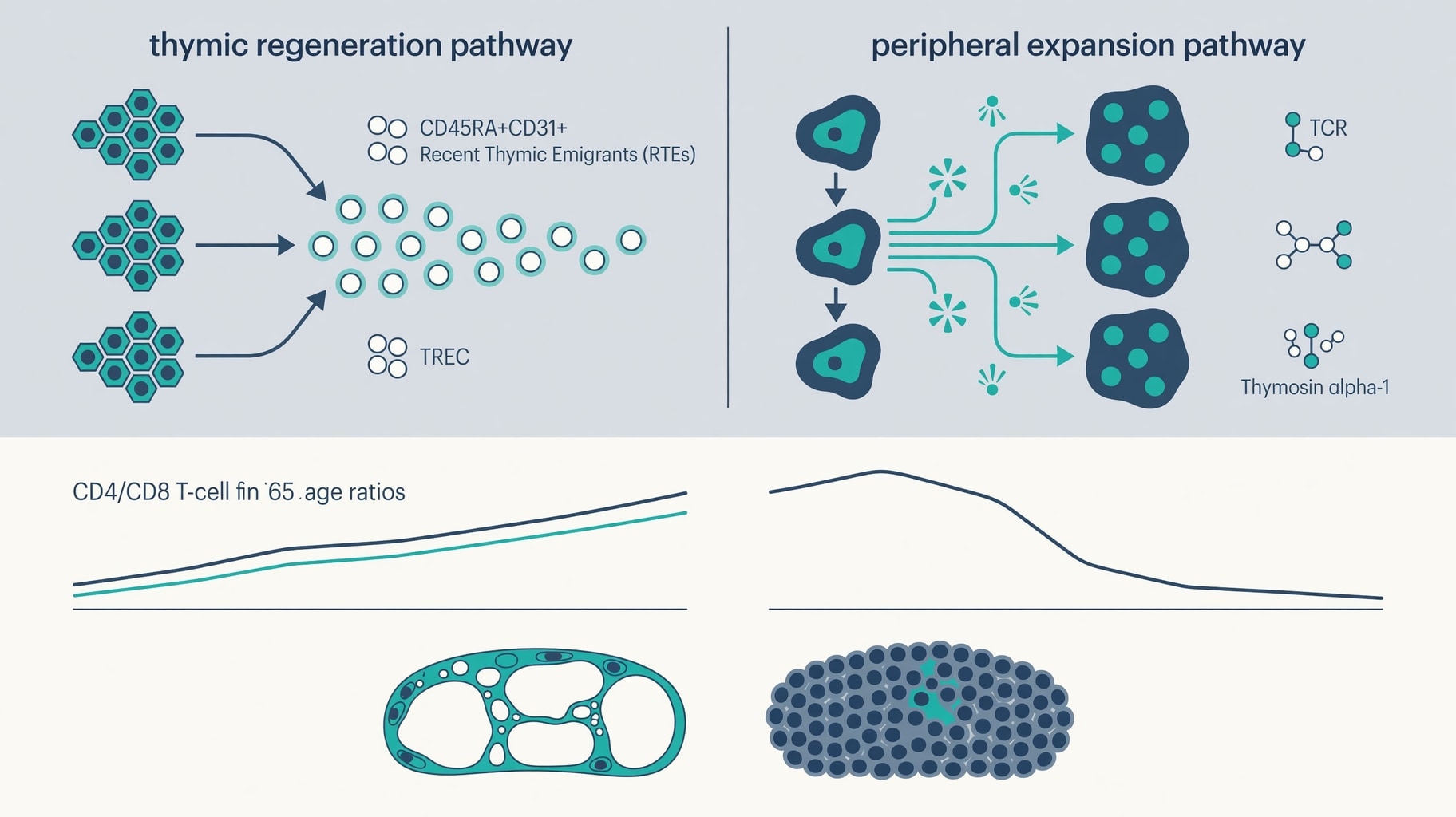
Figure 1: Biological pathways distinguishing thymic regeneration from peripheral T-cell expansion in immunosenescence.
Zinc dependence introduces a biochemical constraint to peptide efficacy that varies significantly between North American and European study populations. Thymulin, a nine-amino acid peptide hormone produced exclusively by thymic reticulo-epithelial cells, requires equimolar binding of zinc to form an active complex. Circulating thymulin levels parallel thymic involution, but unlike the irreversible loss of epithelial structure, thymulin activity is partially restorable through zinc supplementation in deficient elderly individuals according to peptide pharmacology data.
A zinc-deficient patient may fail to respond to thymic peptides not because the epithelium is absent, but because the cofactor necessary for hormone activation is lacking. Spatial ligand-receptor interactions between cortical thymic epithelial cells and thymocytes also decline with age. Recent transcriptomic analyses show reduced Notch pathway activity in circulating T cells from aged donors, reflecting a breakdown in the signaling architecture necessary for lineage commitment as reported in hepatic reconstitution studies.
Peptide interventions that do not address this specific signaling deficit may increase cell numbers without restoring functional repertoire diversity.
Transatlantic regulatory frameworks currently diverge on how to classify these interventions, creating distinct evidentiary hurdles. The European Medicines Agency evaluates many thymic peptides under the Advanced Therapy Medicinal Product framework, requiring demonstration of structural regeneration alongside functional immune recovery. North American regulators frequently categorize similar compounds as dietary supplements or unapproved drugs, where manufacturing consistency often lacks the stringent batch-to-batch validation required for ATMPs.
This regulatory bifurcation means that European trial data increasingly emphasizes histological or imaging confirmation of thymic regrowth, while North American commercial research frequently relies on peripheral blood biomarkers that are less expensive to collect but less specific to thymic function. Researchers comparing datasets across these jurisdictions must account for this methodological asymmetry when synthesizing efficacy claims.
Biomarker Specificity in Trial Endpoints
Total lymphocyte counts are insufficient and potentially misleading markers for thymic rejuvenation because an increase in CD3+ cells following peptide administration could represent benign peripheral expansion of oligoclonal memory populations rather than true restoration of immune competence. Valid assessment of immunosenescence peptide therapy demands cytometry panels capable of identifying recent thymic emigrants.
The combination of CD45RA and CD31 surface markers identifies naive T cells that have recently exited the thymus, distinguishing them from peripherally induced naive-like cells that lack thymic origin according to biomarker validation protocols. Signal joint T-cell receptor excision circles provide a molecular record of thymic output independent of cell surface phenotype. These DNA byproducts of TCR rearrangement are diluted with each peripheral cell division, making them a quantitative measure of new thymic emigrants.
Protocols reviewing thymalin administration have documented cases where undetectable baseline TRECs became measurable only after 16 to 24 weeks of sustained treatment. This timeline suggests thymic reactivation occurs on scales significantly longer than standard 12-week research windows, challenging the design of typical commercial pilot studies that may terminate before biological effects manifest.
Regulatory T-cell phenotyping offers additional resolution for evaluating thymic function. Total Treg percentages often remain stable during peptide intervention, masking underlying changes in thymic versus peripheral tolerance mechanisms. Flow cytometry distinguishing Helios-positive thymic Tregs from Helios-negative peripherally induced Tregs can reveal genuine thymic restoration even when aggregate frequencies appear unchanged as noted in biomarker guidance. Thymic Treg generation depends on functional thymic epithelial cells expressing the autoimmune regulator protein.
These are the same cells targeted by regenerative peptides, making Helios+ Tregs a direct functional readout of target engagement. Imaging biomarkers provide structural confirmation but carry interpretive risks. The TRIIM study demonstrated thymic regrowth on MRI following growth hormone and metformin administration, yet the sample size of nine subjects precludes generalizable conclusions about clinical efficacy.
Increased thymic tissue density on MRI does not definitively confirm functional epithelial regeneration versus reduced adiposity or fibrotic remodeling (ClinicalTrials.gov). Structural volume and functional output are correlated but distinct variables, necessitating that future trials pair imaging with functional cytometry to validate that anatomical changes translate to immunological competence.
FDA guidance on immunogenicity assessments for peptide products further complicates biomarker selection for North American trials. Regulators require demonstration that therapeutic peptides do not induce anti-drug antibodies that could neutralize endogenous thymic hormones or cross-react with host tissues. This safety mandate means that North American protocols must allocate significant resources to immunogenicity monitoring, potentially reducing the statistical power available for efficacy endpoints compared to European studies where some thymic peptides have established safety profiles.
Harmonizing these requirements remains a prerequisite for multinational trials that could adequately power studies on thymic regeneration.
Mechanistic Stratification Beyond Thymic Restoration
The accumulation of GZMK+ effector memory T cells has emerged as a complementary biomarker for tracking immunosenescence velocity alongside naive T-cell restoration. These cells represent a transitional state between functional memory and terminal exhaustion, and their frequency correlates with persistent inflammation and age-linked diseases according to a 2025 Frontiers in Immunology review. Successful thymic restoration should theoretically reduce the relative proportion of this subset by diluting the aged peripheral pool with new naive emigrants.
Monitoring GZMK+ TEM dynamics alongside naive T-cell markers provides a bidirectional assessment of immune age that captures both regenerative input and senescent burden. Mechanistic distinctions extend beyond thymic peptides to other immune-modulating compounds in development. Understanding selank peptide pharmacology reveals alternative pathways for immune modulation that operate through GABAergic and enkephalinergic systems rather than direct thymic stimulation, offering a distinct approach to neuroimmune regulation that does not depend on residual thymic tissue.
Research into kpv peptide mechanism highlights alpha-MSH mediated NF-κB inhibition as a strategy for reducing systemic inflammaging without necessarily rebuilding thymic architecture. An agent that successfully lowers inflammatory markers via KPV may improve healthspan without altering sjTREC levels or naive T-cell counts. Conflating these endpoints would lead to false negatives for anti-inflammatory peptides and false positives for thymic regenerative agents. Mitochondrial approaches also intersect with immune aging through distinct bioenergetic pathways.
Mots c peptide science explores AMPK activation as a means to improve cellular bioenergetics in aged lymphocytes, addressing the metabolic dysfunction that accompanies immunosenescence independently of thymic output. These parallel mechanisms demonstrate that while thymic involution is a central driver of immune aging, it is not the sole determinant of immune function in older adults.
The Thymus Regeneration, Immunorestoration, and Insulin Mitigation extension trial (NCT04375657) currently evaluates biomarkers for epigenetic aging and immunosenescence alongside established clinical measures for prevention of physical frailty, cancer, cardiovascular disease, and infectious diseases including influenza and SARS-CoV-2. This ongoing study represents one of the few registered trials attempting to integrate thymic structural endpoints with broad organismal aging metrics in a controlled setting.
Until such trials report validated outcomes linking sjTRECs and CD45RA+CD31+ recent thymic emigrants to hard clinical endpoints, claims of immune reversal remain mechanistically plausible but clinically unverified.
Further Clinical & Regulatory Context
For deeper analysis and cross-referenced evidence, see: - Related Clinical & Pharmacological Analysis: Aviptadil in ARDS: Divergent Trial Endpoints and Alveolar Type II Signaling - Related Clinical & Pharmacological Analysis: Neoantigen Peptide Vaccines: Prediction Accuracy Versus Clinical T-Cell Expansion