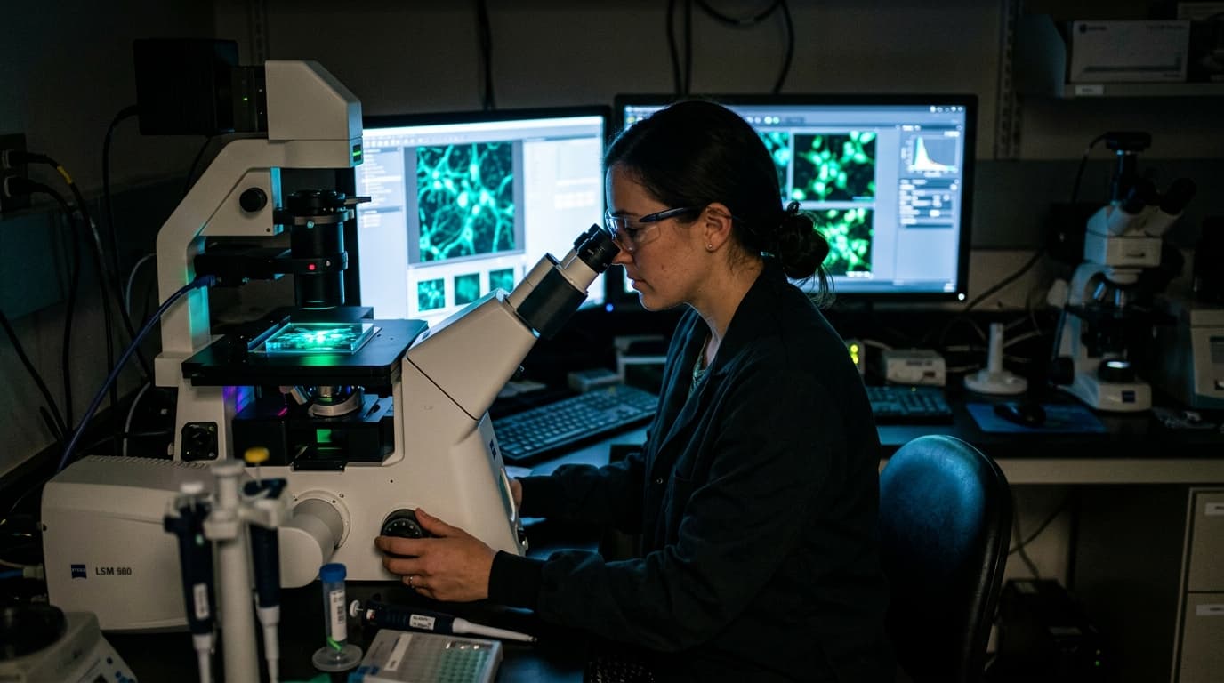
Wash-free imaging of programmed cell death protein 1 (PD-1) in activated T cells was achieved using a library of multicolour fluorogenic amino acids (FgAAs) that also identified avasimibe as a small-molecule modulator in live-cell bioassays. Published September 1, 2026, in Nature Chemistry, this preclinical methodology converts constitutively bright fluorophores into turn-on probes through intramolecular tryptophan-induced quenching. The approach addresses signal-to-noise limitations inherent to conventional peptide labeling by decoupling signal generation from physical washing steps.
This eliminates the need to remove unbound ligands, a process that perturbs live-cell physiology and restricts temporal resolution in high-content screening workflows.
Tryptophan-Induced Quenching Mechanism
Computational analysis using density functional theory confirmed that tryptophan residues positioned adjacent to fluorophore scaffolds suppress emission via photoinduced electron transfer or twisted intramolecular charge transfer until target binding occurs. This environmental sensitivity allows the probe to remain dark in solution and activate only upon specific molecular recognition. The quenching mechanism proved adaptable across ultraviolet to near-infrared fluorophore scaffolds without requiring bespoke synthetic routes for each color variant.
The standardized approach permits integration into solid-phase peptide synthesis workflows. This compatibility with established manufacturing protocols distinguishes the method from specialized labeling techniques requiring genetic encoding. Researchers can generate turn-on probes for flow cytometry and microscopy using routine automated synthesizers.
Validation Distinctions and Photostability Limits
Biological validation focused on PD-1 to demonstrate utility in distinguishing functional receptor states. Fluorogenic peptides targeting PD-1 demonstrated selective activation in activated T cells compared to non-activated controls under no-wash conditions. The investigators applied these wash-free bioassays to screen for small-molecule modulators of PD-1 activity. Avasimibe emerged as a hit compound in the live-cell assay. This finding serves as a proof-of-concept for peptide-enabled drug discovery rather than clinical validation.
The compound requires independent verification in animal models and controlled human trials to establish therapeutic relevance. Cell-based screens identify candidates but cannot predict pharmacokinetics or efficacy in complex biological systems.
This advancement concerns diagnostic tools rather than therapeutic agents. Fluorogenic amino acids serve as molecular reporters, not ligands intended to elicit biological responses. Confusion between imaging probes and bioactive peptides remains common, yet the regulatory and development pathways diverge completely. Therapeutic peptides like dipeptide 2 eye contour formulations target capillary permeability through distinct mechanisms. Similarly, ghk cu copper peptide science focuses on tissue remodeling pathways unrelated to fluorescence reporting.
Research-grade imaging tools also differ from signal peptides such as those studied in matrixyl collagen synthesis protocols. Fluorogenic probes merely report binding events without altering downstream signaling cascades.
The current evidence base derives entirely from in vitro cell culture systems. No data exists regarding probe stability, biodistribution, or toxicity in living organisms. Translation to in vivo imaging would require extensive pharmacokinetic characterization absent from the current publication. Supplementary data indicates that while Si-rhodamine FgAA peptides enabled wash-free imaging with minimal background, extended illumination periods risk photobleaching and cellular stress.
Researchers must now quantify phototoxicity thresholds to determine whether these tools permit multi-hour time-lapse studies without compromising T cell viability.