Endorphins vs. Enkephalins: Biochemistry and Pain Modulation Explained
Beta-endorphin is a 31-amino-acid peptide derived from the proopiomelanocortin (POMC) precursor with high affinity for mu-opioid receptors, whereas enkephalins are pentapeptides derived from proenkephalin that act primarily as delta-opioid receptor agonists. This fundamental distinction in endorphins vs enkephalins biochemistry determines their physiological roles far more than cultural associations with wellness or exercise. While consumer health content in both English and Spanish often treats these molecules as interchangeable "feel-good" chemicals, biochemical records demonstrate they are distinct molecular families with separate genetic origins, enzymatic processing pathways, and receptor selectivities that produce divergent downstream effects.
The functional divergence begins at the gene level and extends through every stage of biological activity. Beta-endorphin’s amphipathic alpha-helical structure confers a significantly longer plasma half-life than the rapidly degraded enkephalin pentapeptides, according to structural analyses documented by Peptide Insight. Furthermore, their receptor preferences produce fundamentally different signaling outcomes: mu-opioid activation by beta-endorphin mediates analgesia and euphoria, while delta-opioid activation by enkephalins modulates mood and anxiety with less abuse potential, as detailed in receptor binding reviews by Rethink Peptides. Clinical evidence using opioid blockade now challenges the pervasive attribution of the runner’s high solely to endorphins, as circulating beta-endorphin does not readily cross the blood-brain barrier and naltrexone administration does not consistently abolish exercise-induced euphoria.
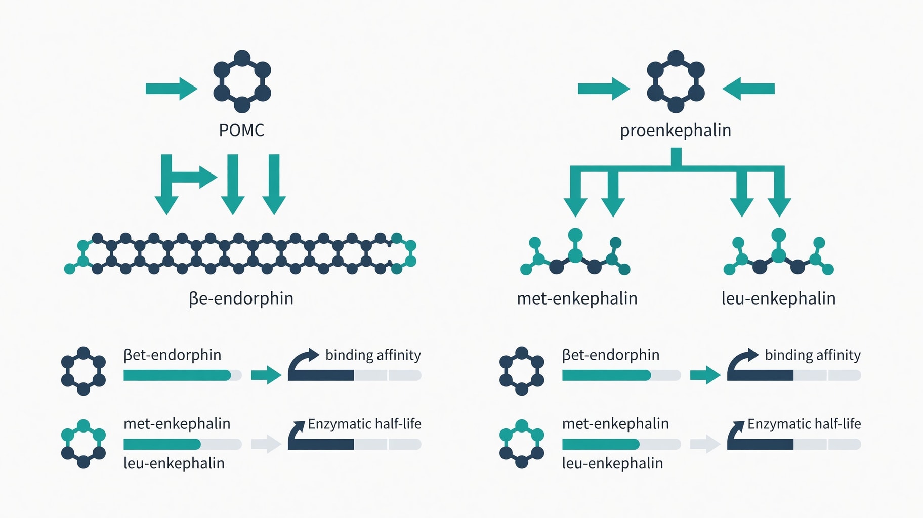
Figure 1: Biochemical divergence between beta-endorphin (POMC-derived, mu-opioid selective) and enkephalins (proenkephalin-derived, delta-opioid selective) showing precursor processing, receptor affinity, and functional outcomes.
Precursor Processing and Structural Divergence
The biosynthesis of endogenous opioids follows strictly regulated enzymatic cascades that dictate which peptides emerge from each precursor protein. Beta-endorphin originates from the 241-amino-acid POMC precursor, encoded by the POMC gene on chromosome 2p23.3. Tissue-specific processing by prohormone convertases PC1/3 and PC2 cleaves this precursor to yield beta-endorphin alongside adrenocorticotropic hormone (ACTH) and melanocyte-stimulating hormones. This shared precursor explains why stress-induced ACTH release often coincides with elevated beta-endorphin levels in circulation. The processing cascade is tightly regulated, meaning beta-endorphin production cannot be isolated from other POMC-derived peptides in physiological contexts.
Enkephalins derive from an entirely separate genetic source. The proenkephalin (PENK) gene encodes a precursor containing four copies of Met-enkephalin, one copy of Leu-enkephalin, and two extended enkephalin sequences. Enzymatic cleavage of proenkephalin yields these pentapeptides independently of the POMC system. This genetic separation means that physiological states or interventions affecting POMC processing will not necessarily alter enkephalin levels, and vice versa. The PENK gene product is widely distributed throughout the central and peripheral nervous systems, whereas POMC-derived beta-endorphin is primarily synthesized in the anterior pituitary and arcuate nucleus of the hypothalamus (Mdpi).
Structural differences between these peptide families directly influence their stability and bioavailability. Beta-endorphin adopts an amphipathic alpha-helical conformation in its C-terminal region, approximately residues 13 through 31. This secondary structure protects the peptide from rapid enzymatic degradation and accounts for its considerably longer biological half-life compared to enkephalins. The pentapeptide structure of enkephalins, lacking this protective helical domain, renders them susceptible to rapid breakdown by enkephalinases and other peptidases in plasma and synaptic clefts. Consequently, enkephalins function as fast-acting, spatially restricted neuromodulators, while beta-endorphin can exert more sustained hormonal effects.
Both families share a common N-terminal Tyr-Gly-Gly-Phe motif, known as the opioid message domain, which is responsible for receptor recognition. However, the C-terminal address domain differs substantially between beta-endorphin and enkephalins. This address sequence determines receptor selectivity and binding affinity. The extended C-terminus of beta-endorphin confers high affinity for both mu and delta receptors, though with preference for mu. The shorter enkephalin C-termini favor delta-opioid receptor binding. These structural distinctions mean that even when both peptides are present in the same neural circuit, they activate different receptor populations and trigger distinct intracellular signaling cascades.
Receptor Selectivity and Signaling Outcomes
Opioid receptors are G protein-coupled receptors designated mu, delta, and kappa, each producing distinct physiological effects when activated. Beta-endorphin serves as the most potent endogenous ligand for the mu-opioid receptor. Mu-receptor activation produces analgesia, euphoria, respiratory depression, and physical dependence. This receptor subtype is the primary target of clinical pain management with pharmaceutical opioids. Beta-endorphin also binds delta receptors with significant affinity, but its mu selectivity dominates its physiological profile. The peptide’s role in stress-induced analgesia and reward circuitry stems primarily from mu-receptor activation in the ventral tegmental area and nucleus accumbens.
Enkephalins act preferentially as agonists at the delta-opioid receptor, with secondary affinity for mu receptors. Delta-receptor activation modulates mood, anxiety, and moderate pain transmission through presynaptic inhibition in the spinal cord dorsal horn. Unlike mu-receptor agonists, delta agonists produce anxiolytic and antidepressant-like effects in animal models with substantially less abuse potential. This selectivity profile makes enkephalins functionally distinct from beta-endorphin despite their shared classification as endogenous opioids. Delta receptors predominate in the basal ganglia and limbic systems, positioning enkephalins as key regulators of emotional processing rather than pure analgesia.
The distinction extends to therapeutic implications. Research into non-pharmacological pain modulation, including manual therapy and acupuncture, suggests frequency-dependent effects on endogenous opioid release. Low-frequency electroacupuncture at 2 Hz preferentially stimulates release of beta-endorphin, Met-enkephalin, and endomorphin, activating both mu and delta receptors. However, the analgesic effects attributed to such interventions may result from centrally synthesized peptides independent of peripheral measurements, or from non-opioid mechanisms producing similar subjective effects. The complexity of these pathways means that measuring plasma beta-endorphin alone cannot confirm central opioid-mediated analgesia.
Pharmacological agents targeting these systems face challenges due to rapid peptide degradation. Enkephalinase inhibitors like thiorphan were developed to prolong enkephalin activity by blocking enzymatic breakdown. Similarly, selank peptide pharmacology has been investigated for potential modulation of enkephalin degradation pathways, though clinical validation remains limited. These approaches aim to enhance endogenous opioid tone rather than replace it with exogenous agonists. The therapeutic window is narrow, as excessive delta-receptor activation can produce tolerance and cross-tolerance with mu receptors. Understanding the precise receptor selectivity of each endogenous peptide is essential for developing targeted interventions that avoid the dependence liability associated with non-selective mu activation.
For researchers examining inflammatory modulation pathways, the distinction between opioid and non-opioid peptide systems matters. The kpv peptide mechanism operates through alpha-MSH receptor pathways rather than opioid receptors, despite alpha-MSH also deriving from the POMC precursor. This illustrates how a single precursor can yield peptides with entirely different receptor targets and physiological functions. Similarly, klow blend peptide review literature highlights combinations targeting inflammation through non-opioid mechanisms, underscoring that pain and mood modulation involve multiple peptide families beyond the classical opioid system.
Reevaluating Exercise-Induced Euphoria and Clinical Translation
The cultural narrative of the runner’s high as a purely endorphin-mediated phenomenon has faced significant challenge from clinical blockade studies. Circulating beta-endorphin cannot readily cross the blood-brain barrier, raising questions about how peripheral elevations during exercise could produce central euphoria. Research published in PNAS demonstrated that wheel running raises both beta-endorphin and anandamide in mice, but genetic and pharmacological interruption of cannabinoid signaling abolished the anxiolytic effects, while opioid blockade did not consistently prevent euphoria. Human trials using naltrexone, a mu-opioid receptor antagonist, found that the share of participants reporting subjective runner’s high was comparable between naltrexone and placebo groups.
This evidence does not demonstrate that opioids contribute nothing to exercise-induced mood changes. Naltrexone did not erase every chemical response to running, and studies have focused primarily on euphoria and anxiety rather than all forms of pain relief or stress regulation. The findings represent evidence against necessity, not evidence of total absence. Central opioid activity can accompany a high, but it may not be required for the subjective experience. Endocannabinoids like anandamide and 2-AG are lipid signaling molecules that interact with CB1 and CB2 receptors and can cross the blood-brain barrier more readily than peptide opioids. Current models suggest the runner’s high involves an orchestra of interacting brain systems rather than a single molecular pathway.
The persistence of the endorphin myth in bilingual wellness communities reflects broader challenges in translating biochemistry into accessible health information. Spanish-language content often uses "endorfinas" as a catch-all term for exercise benefits, mirroring English-language oversimplifications. Yet the biochemical records show that attributing all post-exercise mood enhancement to beta-endorphin ignores the documented roles of endocannabinoids, enkephalins, and non-opioid neuromodulators. This matters for consumers evaluating products or protocols marketed as endorphin boosters. If the desired effect depends on central mechanisms, interventions that only elevate peripheral beta-endorphin may not produce the promised psychological benefits.
Clinical research continues to investigate how endogenous opioid systems can be modulated for pain management without the risks of exogenous opioids. Trials registered on ClinicalTrials.gov explore interventions ranging from manual therapy to novel enzyme inhibitors. However, human CNS binding kinetics for endogenous peptides remain incompletely mapped, and most receptor affinity data derive from in vitro or animal models. The gap between preclinical pharmacology and clinical outcomes remains substantial. Patients and practitioners should interpret claims about endorphin or enkephalin modulation with awareness of these evidence limitations.
The biochemical distinction between endorphins and enkephalins is well-established through decades of molecular characterization. Their precursor genes, processing enzymes, receptor selectivities, and degradation pathways are documented in peer-reviewed literature. What remains uncertain is how reliably these molecular mechanisms translate to predictable subjective experiences in humans. The runner’s high research exemplifies this gap: robust biochemistry coexists with inconsistent clinical blockade data. Future progress depends on studies that measure central peptide activity directly rather than inferring it from peripheral markers or subjective reports. Until such evidence emerges, the functional differences between these peptide families should be understood as biochemical facts distinct from their cultural associations.
Further Clinical & Regulatory Context
For deeper analysis and cross-referenced evidence, see: - Related Clinical & Pharmacological Analysis: Cholecystokinin Satiety Signaling Depends on Vagal Afferents, Not Willpower