
Structural characterization studies establish that elastin-derived peptides containing the VGVAPG sequence exhibit optimal chemotactic activity for fibroblasts at approximately 10⁻⁸ M, with binding dependent on a type VIII β-turn conformation of the GxxPG motif that interacts specifically with the elastin-binding protein subunit of the elastin receptor complex. This precise molar threshold separates the hexapeptide from non-specific elastin hydrolysates by triggering discrete intracellular signaling cascades via the 67-kDa EBP rather than passive accumulation.
The interaction upregulates matrix metalloproteinase-2 and MMP-3 expression while simultaneously directing cell migration, creating a regulated feedback loop between extracellular matrix remodeling and cellular response. For regulatory scientists evaluating structure-function claims, these defined kinetic parameters provide measurable biological endpoints distinct from generic structural protein degradation.
VGVAPG functions as a specific ligand rather than a passive breakdown product, a classification supported by receptor binding assays and downstream transcriptional data. Research published in Nature Aging demonstrates that while circulating elastin fragments increase with age and drive systemic inflammation through innate immune activation, the specific hexapeptide sequence operates through a distinct receptor-mediated mechanism that can be isolated in vitro (Nature Aging).
The peptide does not merely replace lost elastin mass; it actively engages a heterotrimeric receptor complex to modulate proteolytic enzyme expression and direct cellular movement. This dual function positions VGVAPG as a signaling molecule with defined downstream consequences in dermal biology, separate from the bulk material properties of elastin itself. Evaluating topical applications containing this sequence therefore requires assessing receptor engagement and enzymatic modulation rather than simple moisturization or protein supplementation.
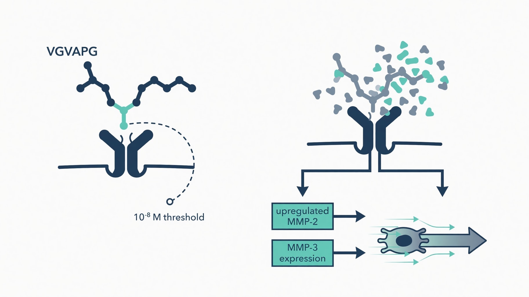
Figure 1: VGVAPG-ERC binding kinetics and downstream fibroblast chemotaxis signaling pathway.
ERC Binding Kinetics and Structural Requirements
Bioactivity requires more than the presence of the six-amino-acid sequence; the peptide must adopt a specific three-dimensional structure to engage its receptor. Structural analyses confirm that the GxxPG motif within VGVAPG must form a type VIII β-turn conformation to bind the elastin-binding protein, a constraint that explains why linear peptide analogs or denatured elastin hydrolysates often fail to replicate the signaling potency of the native sequence (Frontiers in Cardiovascular Medicine).
The turn structure presents the valine and proline residues in a spatial orientation that fits the binding pocket of the EBP, rendering the peptide biologically inert if this geometry is disrupted regardless of its primary sequence. Molecular modeling and site-directed mutagenesis have identified the exact residues on the receptor responsible for this interaction.
Research published in PubMed identified Gln-97 and Asp-98 as essential residues for VGVAPG binding because they define the elastin-binding pocket on the EBP, while additional residues including Leu-103, Arg-107, and Glu-137 interact directly with the peptide itself (PubMed). Mutagenesis experiments at these positions validated the structural model, providing atomic-level data concerning the VGVAPG-EBP interaction that allows researchers to distinguish true receptor ligands from non-specific binding events in assay validation.
The elastin receptor complex itself is a heterotrimer composed of three distinct proteins, and signal transduction requires the assembly of all subunits. The EBP serves as the peripheral membrane protein that directly binds the peptide, but functional signaling depends on protective protein/cathepsin A and neuraminidase-1. Coverage in Frontiers in Cardiovascular Medicine notes that neuraminidase-1 is essential for ERC signal transduction and activates downstream pathways that vary significantly among cell types (Frontiers in Cardiovascular Medicine).
This complexity means that VGVAPG binding alone is insufficient for biological effect; the complete receptor complex must be assembled and functional on the cell surface. Formulations targeting this pathway must therefore consider receptor density and subunit availability alongside peptide concentration. Binding affinity measurements further define the operational window for this interaction.
In 48-microwell chemotaxis chambers, the peptide demonstrates maximal activity within a narrow nanomolar range, and concentrations significantly above or below this optimum show reduced efficacy.
This bell-shaped dose-response curve is characteristic of receptor-mediated signaling and contrasts with the linear responses typically seen with non-specific chemotactic factors. The saturation kinetics suggest that VGVAPG occupies a finite number of high-affinity binding sites, and excess peptide may induce receptor desensitization or internalization, effectively shutting down the signaling cascade. This kinetic behavior has direct implications for formulation design, where higher concentrations do not necessarily yield greater biological responses and may instead inhibit activity.
Proteolytic Signaling and Matrix Metalloproteinase Regulation
Receptor engagement triggers measurable changes in proteolytic enzyme expression as an active transcriptional response rather than a passive degradation process. Studies on melanoma invasion demonstrate that VGVAPG enhances the expression of the elastin-degrading enzymes MMP-2 and MMP-3, amplifying the invasive capacity of cells by upregulating these specific matrix metalloproteinases (PubMed).
This finding establishes a direct mechanistic link between elastin fragment signaling and extracellular matrix remodeling, where the peptide acts as a feedback signal informing cells of elastin degradation status and triggering compensatory or pathological proteolytic responses. The proteolytic response extends beyond MMP-2 and MMP-3 to include broader homeostatic regulation.
Research on mouse cortical glial cells shows that VGVAPG modulates the expression of MMP-2, MMP-9, and multiple tissue inhibitors of metalloproteinases, suggesting a sophisticated mechanism that recalibrates the proteolytic balance rather than simply promoting degradation (PubMed). This nuanced response is critical for understanding the peptide's role in tissue remodeling versus pathological destruction; in aging skin, the balance may shift toward net degradation, but the signaling mechanism itself remains a regulated cellular response.
Endothelial cells exhibit a distinct proteolytic profile upon VGVAPG exposure, reinforcing the concept that signaling is context-dependent. Investigations into angiogenesis reveal that the hexapeptide upregulates MT1-MMP, a membrane-type metalloproteinase essential for endothelial tubulogenesis and migration (PubMed). The same peptide sequence elicits different enzymatic responses in fibroblasts, melanoma cells, glial cells, and endothelial cells, meaning formulators cannot assume a universal proteolytic outcome across all dermal cell populations.
The local cellular composition of the target tissue will determine the specific enzymatic response to the peptide. The generation of VGVAPG itself is tightly linked to specific proteolytic enzymes, creating a self-reinforcing cycle. Macrophage-derived MMP-12 can completely degrade tropoelastin within 30 minutes, releasing bioactive peptides enriched in VGVAPG motifs.
Reporting in eLife describes this as a feed-forward positive feedback loop where elastin degradation generates peptides that signal through the ERC to reinforce inflammatory activation and proteolytic responses (eLife).
In chronic inflammatory conditions or advanced photoaging, this loop may become dysregulated. Understanding this amplification mechanism is essential for distinguishing therapeutic remodeling from pathological degradation, as the peptide is both a product of proteolysis and a regulator of future proteolytic activity.
Cell-Type Specific Chemotaxis Thresholds and Safety Parameters
Chemotactic potency varies dramatically across cell types, defying generalized efficacy claims and necessitating cell-specific safety assessments. For dermal fibroblasts and monocytes, optimal chemotaxis occurs at approximately 10⁻⁸ M. Structural characterization research confirms that this activity is substantial, reaching half or greater of the maximum response to platelet-derived growth factor for fibroblasts (ResearchGate). This benchmark establishes VGVAPG as a physiologically relevant chemoattractant in connective tissue remodeling contexts.
Tumor cells display markedly different sensitivity thresholds that raise safety considerations.
Lewis lung carcinoma cells exhibit maximal chemotactic response at 5 nM when assayed in modified Boyden chambers, a nanomolar potency twenty-fold higher than the optimal concentration for normal fibroblasts (PubMed). This discrepancy suggests that malignant transformation may alter ERC expression or coupling efficiency, implying that safety assessments using only normal fibroblasts may underestimate the peptide's bioactivity in transformed cell populations.
Regulatory evaluations must account for these cell-type specific variations in receptor sensitivity to ensure that topical application does not inadvertently promote neoplastic migration.
Keratinocytes present a biphasic response that complicates formulation dosing for topical products. In vitro studies on cultured keratinocytes show that VGVAPG elicits chemotactic responses at 10⁻⁹ M, a full log lower than the optimal fibroblast concentration, yet treatment at 10⁻⁶ M or 10⁻⁵ M suppresses cell growth and increases terminal differentiation markers (PubMed). This narrow therapeutic window means that concentrations effective for fibroblast recruitment may be inhibitory or differentiative for epidermal cells.
Topical formulations targeting the dermis must handle this stratified sensitivity, as overdosing could compromise barrier function while attempting to stimulate dermal repair. Immune cell recruitment represents another dimension of VGVAPG bioactivity with regulatory implications. The peptide promotes T-cell differentiation toward a Th-1 cytokine profile by binding the 67-kDa spliced-galactosidase elastin receptors. Research in the Exploratio Journal indicates that this signaling enhances IFN-γ, IL-2, and IL-12 production while restricting Th-2 cytokines (Exploratio Journal).
This immunomodulatory effect connects dermal application to adaptive immune responses. In compromised or aged skin, this Th-1 polarization could support antimicrobial defense or exacerbate chronic inflammation depending on the existing inflammatory milieu and the integrity of the skin barrier.
Translating these in vitro molar concentrations to topical formulation percentages remains scientifically unjustified without further clinical data. The assays utilize direct exposure in defined buffer systems, accounting for no penetration barriers, metabolic degradation, or receptor desensitization over time. Most structural and kinetic data derive from non-human or immortalized cell lines, limiting direct extrapolation to primary human dermal fibroblasts in vivo.
Furthermore, the biphasic responses observed in keratinocytes suggest that safety margins are narrower than typical cosmetic ingredients.
Pending verification of receptor density in aged human skin and the establishment of clinical dose-response relationships, the optimal topical concentration for VGVAPG remains a parameter requiring empirical determination rather than theoretical calculation from binding kinetics. Regulatory submissions for products containing this peptide must therefore include specific safety data addressing the differential sensitivity of keratinocytes and potential neoplastic cell lines, rather than relying solely on fibroblast proliferation assays.